HOME
ABOUT US
REFERENCES
REPRESENTATIONS
CONTACT US
TÜRKÇE
Engineering and Technical Education
PHYSICS
ELECTRONICS
COMMUNICATIONS
ELECTRICITY
ENERGY
MECHATRONICS, AUTOMATION & COMPUMECHATRONICS
MECHANICS
FLUID MECHANICS
THERMODYNAMICS & THERMOTECHNICS
PROCESS CONTROL
CHEMICAL ENGINEERING
FOOD & WATER TECHNOLOGIES
ENVIRONMENT
BIOMEDICAL ENGINEERING
Physics, Chemical, Biology Education
OPTIKASCIENCE
DATA HARVEST
ERLER-ZIMMER
Maths Education
ERLER-ZIMMER
3D Anatomy Series
1. OPTIKASCIENCE
1.1. Section 01 - Kits
1.1.1. Physics Laboratory Sets
1.1.2. Basic Kits
1.1.3. Advanced Kits
1.2. Section 02 - Physics
1.2.1. Statics of solids
1.2.2. Dynamics
1.2.3. Translational motion
1.2.4. Rotational motion
1.2.5. Oscillatory motion
1.2.6. Inertia- Collisions - Two-dimension motion
1.2.7. Liquids
1.2.8. Gases and vacuum
1.2.9. Wave's propagation
1.2.10. Sound Waves
1.2.11. Molecular aspect of matter
1.2.12. Temperature and Heat
1.2.13. Geometrical Optics
1.2.14. Wave Optics
1.2.15. Optical Benches
1.2.16. Electrostatics
1.2.17. Electrical conduction
1.2.18. Magnetism and electromagnetism
1.2.19. Atomic Physics
1.3. Section 03 - Technique and Energy
1.3.1. Energy conversions
1.3.2. Renewable energies
1.4. Section 04 - Microscopy
1.4.1. On-field microscopy kits
1.4.2. Biological microscopes
1.4.3. Stereomicroscopes
1.4.4. Multimedia system
1.4.5. Microscopy accessories
1.4.6. Optical magnifiers
1.4.7. Prepared slides for microscopy
1.5. Section 05 - Biology
1.5.1. Botany
1.5.2. Zoology
1.5.3. Experiments on human beings
1.5.4. Human anatomy and DNA models
1.6. Section 06 - Ecology
1.6.1. Kit for environmental analysis
1.6.2. Items for sample's collection
1.6.3. Stations for the detection of air pollution
1.6.4. Digital instruments
1.7. Section 07 - Meterology
1.7.1. Instruments and weather stations
1.8. Section 08 - Astronomy and Earth Science
1.8.1. Rocks, fossils and minerals
1.8.2. Geological models
1.8.3. The Earth and the solar system
1.9. Section 09 – Chemistry
1.9.1. Periodic table of elements
1.9.2. Molecular models and atomic models
1.9.3. PH-meters
1.9.4. Refractometry
1.9.5. Polarimetry
1.9.6. Spectroscopy
1.9.7. Electrology
1.10. Section 10 – On-Line Science
1.10.1. Interfaces
1.10.2. MBL Sensors
1.10.3. USB Sensor
1.11. Section 11 – Drawing and Mathematics
1.11.1. Drawing
1.11.2. Enumeration
1.11.3. Logics
1.11.4. Fractions and percentages
1.11.5. Geometry
1.11.6. Mathematics on magnetic blackboard
1.12. Section 12 – Measurement Instruments
1.12.1. Lengths and angles
1.12.2. Volumes/Time intervals
1.12.3. Temperature
1.12.4. Density/Forces, weights and masses
1.12.5. Electrical devices
1.13. Section 13 – Lab Tools
1.13.1. Items and instruments
1.13.2. Electrical power sources
2. DATA HARVEST
2.1. Data Logging
2.1.1. Data Loggers
2.1.1.1. Data Logging Range
2.1.2. Sensors
2.1.2.1. Sensor Range
2.1.3. Apps
2.1.3.1. Cross-platform software
2.1.4. Accesories
2.1.4.1. All Accessories
2.2. Dynamics
2.2.1. Dynamics System
2.2.2. Software
2.2.2.1. Cross-platform software
2.2.3. Accessories
2.2.3.1. All Accessories
2.3. Teaching
2.3.1. Teaching Packs
2.3.2. Teaching Metarials
2.4. More Science
2.4.1. Smart Microscopes
2.4.2. Genecon Hand-Held Dynamo
2.4.3. Renewable Energy
2.5. Product Brochures
2.5.1. Primary Teaching Brochure
2.5.2. Secondary Teaching Brochure
3. ERLER-ZIMMER
3.1. Anatomical Models
3.1.1. Skeleton Models
3.1.1.1. Pelvises
3.1.1.2. Limbs
3.1.1.3. Full Body
3.1.1.4. Skulls
3.1.1.5. Spines
3.1.1.6. Bones
3.1.2. Organs & Structures
3.1.2.1. Respiratory System
3.1.2.2. Brain & Nerves
3.1.2.3. Urinary System
3.1.2.4. Skin-Hair-Nail
3.1.2.5. Head Models
3.1.2.6. Circulatory System - Blood Vessels
3.1.2.7. Circulatory System - Hearts
3.1.2.8. Circulatory System - Circulation
3.1.2.9. Liver
3.1.2.10. Muscular Models
3.1.2.11. Kidneys
3.1.2.12. Ears
3.1.2.13. Torsos
3.1.2.14. Digestive System
3.1.2.15. Teeth
3.2. 3D Anatomy Series
3.3. Medical Simulators
3.3.1. Auscultation
3.3.2. Endoscopy
3.3.3. Gynaecology
3.3.4. Skin Suture
3.3.5. Injection
3.3.6. Nursing
3.3.6.1. Chronic Wounds
3.3.6.2. Diabetes
3.3.6.3. Catheterisation
3.3.6.4. Nursing Dolls - Baby & Child Dolls
3.3.6.5. Nursing Dolls - Adult Dolls
3.3.6.6. Stoma Care
3.3.7. Cancer Prevention
3.3.8. Emergency
3.3.8.1. Airway
3.3.8.2. CPR - Baby & Child Dolls
3.3.8.3. CPR - Adult Dolls
3.3.8.4. All Dolls - Baby & Child Dolls
3.3.8.5. All Dolls - Adult Dolls
3.3.8.6. Rescue
3.3.8.7. Thorax
3.3.8.8. Trauma Simulation
3.3.9. X-RAY / CT
3.3.10. Pregnancy & Birth
3.3.11. Ultrasound
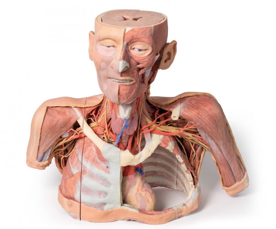
Head, Neck and Shoulder with angiosomes
This large 3D printed specimen displays a great deal of anatomy spanning the head, neck, thorax, axillae and upper limbs. Detailed anatomical description on request....
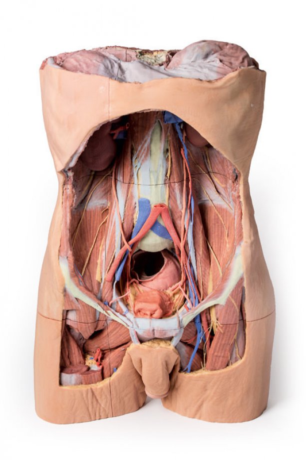
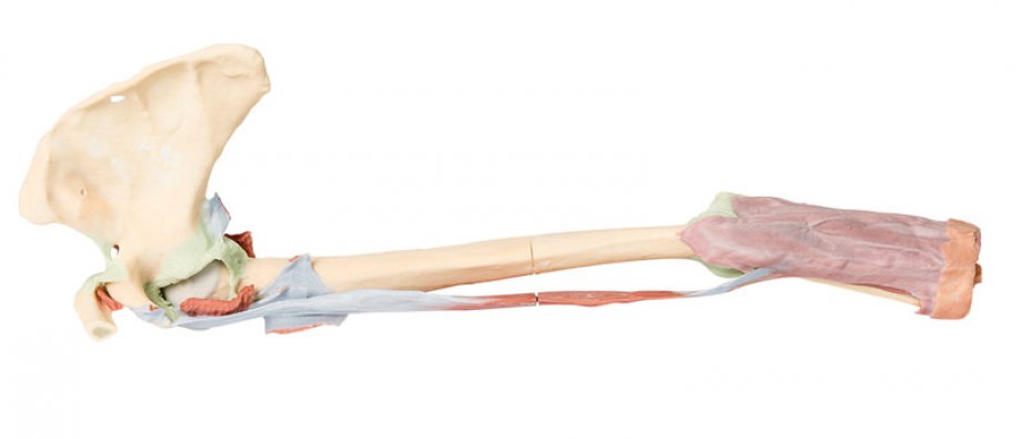
Posterior Abdominal wall
This large 3D-printed specimen displays the entire male posterior abdominal wall from the diaphragm to the pelvic brim, as well as pelvic anatomy and to the proximal thigh. This same individual specimen is also available as a pelvic and proximal thigh specimen (MP1770). Detailed anatomical description on request....
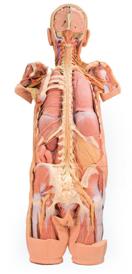
Nervous System Dissection (posterior view)
This 3D printed specimen presents a unique view of axial anatomy, presenting a dorsal deep dissection of the head, neck, axillae, thorax, abdomen, and gluteal regions. The removal of the posterior portions of the cranium and laminectomy from the cervical region to the opening of the sacral canal affords a continuous view of the central nervous system structures and origin of the segmental nerves relative to other axillary and appendicular structures. Detailed anatomical description on request....
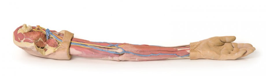
Upper Limb
This 3D-printed specimen demonstrates the superficial anatomy of a left upper limb from the blade of the scapula to the hand. The skin and superficial and deep fascia has been removed from most of the limb except over the dorsum of the scapula, proximal arm, and over the hand. The superficial veins, including the median cubital vein, have been maintained; with the cephalic and basilica preserved from the wrist to the deltopectoral groove and termination in the brachial vein, respectively. Detailed anatomical description on request....
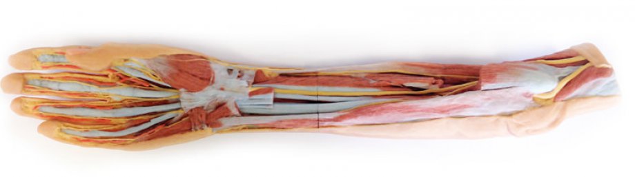
Forearm and hand - superficial and deep dissection
This 3D printed specimen preserves a mixed superficial and deep dissection of the anterior aspect of a right distal arm, forearm and hand. Detailed anatomical description on request....
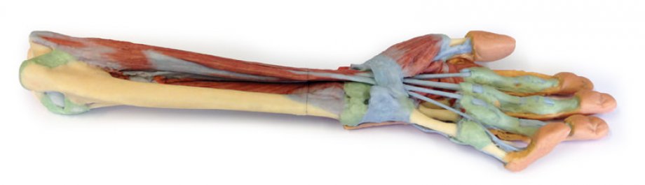
Forearm and hand - deep dissection
This 3D printed specimen of a left upper limb preserves a deep dissection from the distal humerus to the palmar surface of hand. Detailed anatomical description on request....
Upper Limb - biceps, bones and ligaments
This 3D-printed specimen shows the origin and insertion of biceps (most other arm and shoulder muscle bellies have been removed). The long head of biceps arises from the supraglenoid tubercle (hidden from view) and travels inferiorly in the bicipital groove, whereas the short head of biceps arises from the coracoid process. The bifid insertion of the muscle as the bicipital aponeurosis and the rounded tendon which can be seen winding around the radius to insert into the radial tuberosity are clearly discernable. Detailed anatomical description on request....
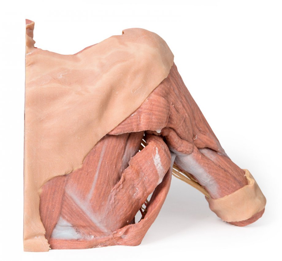
Right thoracic wall - axilla, and the root of the neck
This 3D printed specimen preserves a dissection of the right thoracic wall, axilla, and the root of the neck. The specimen is cut just parasagittally and the visceral contents of the chest have been removed. Structures within the right chest wall are visible deep to the parietal pleura, including the ribs, muscles of the intercostal spaces and the origins of the neurovascular bundle in each intercostal space. The pectoralis major has been reflected medially towards the sectioned edge of the specimen to expose pectoralis minor which acts as a useful landmark as it divides the axillaryartery into its three parts. T...
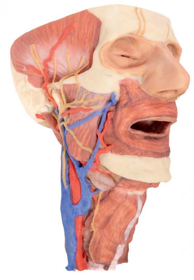
Head and visceral column of the neck
This 3D print specimen preserves a series of features of the head and visceral column of the neck: The face: On the right side of the head the parotid gland has been removed to reveal the facial nerve and all its branches (temporal, zygomatic, buccal, marginal mandibular and cervical) and demonstrate the spatial relations of structures embedded in the gland from superficial to deep (facial nerve, retromandibular vein, external carotid artery). In the surrounding region the temporalis, masseter and posterior belly of digastric are exposed, as are and the facial artery, transverse facial artery and superficial tem...
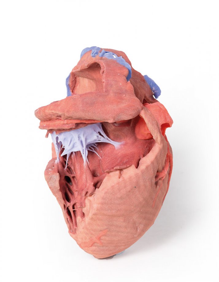
Heart internal structures
This 3D printed heart has been dissected to display the internal structures of the chambers. At the base of the heart the termination of the superior vena cava is preserved entering the right atrium. Part of the inferior vena cava is also preserved on the inferior aspect of the right atrium; however, most of the vessel lumen and much of the anterior wall has been removed to expose the pectinate muscles of the right auricle and the fossa ovalis (which is nearly translucent in the 3D print). The anterior wall of the right ventricle has also been removed to expose the right atrioventricular valve and its three cusps...