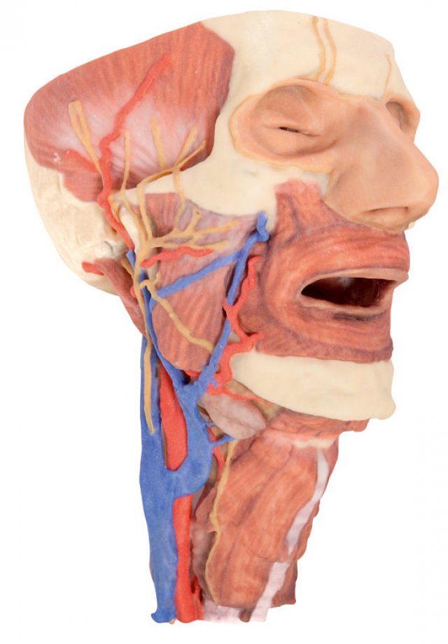
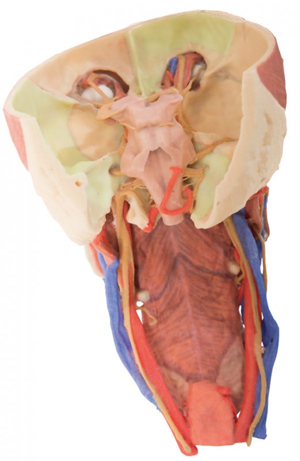
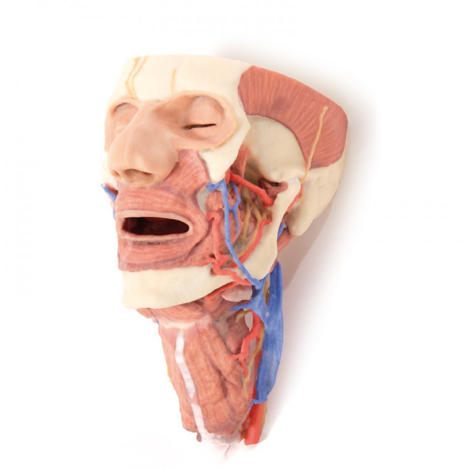
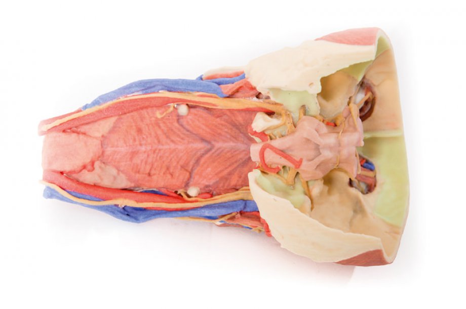
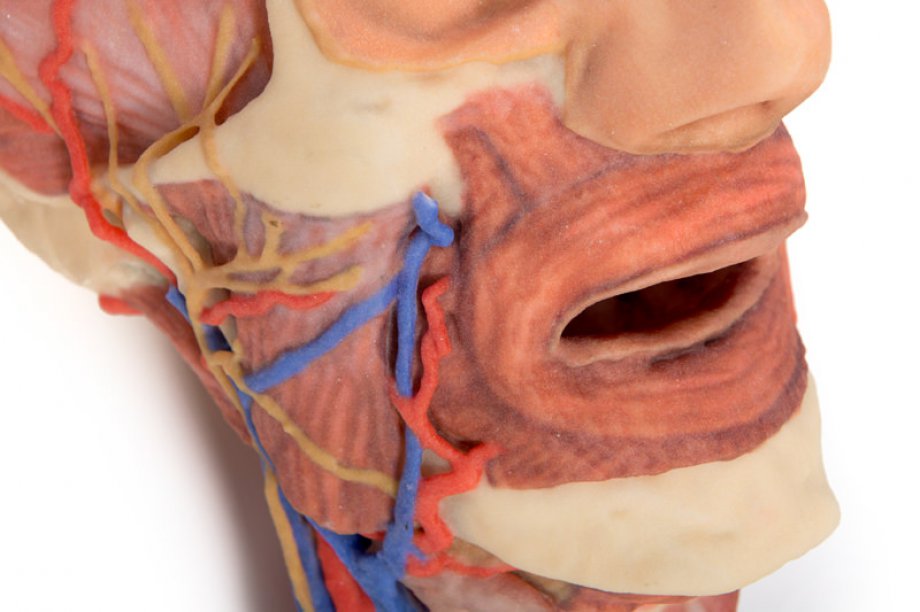
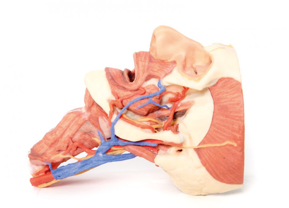
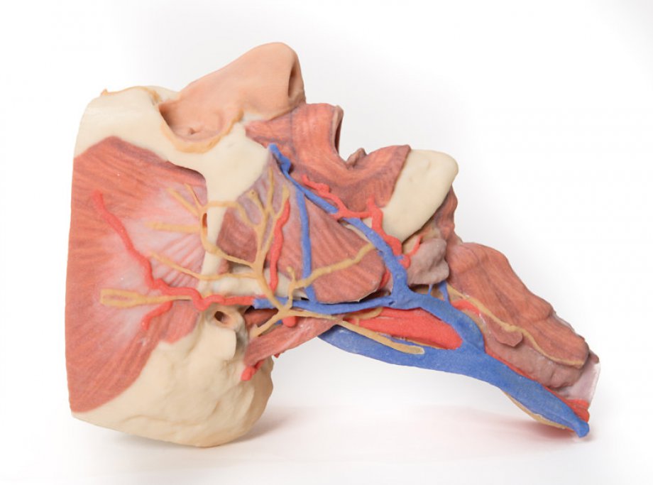
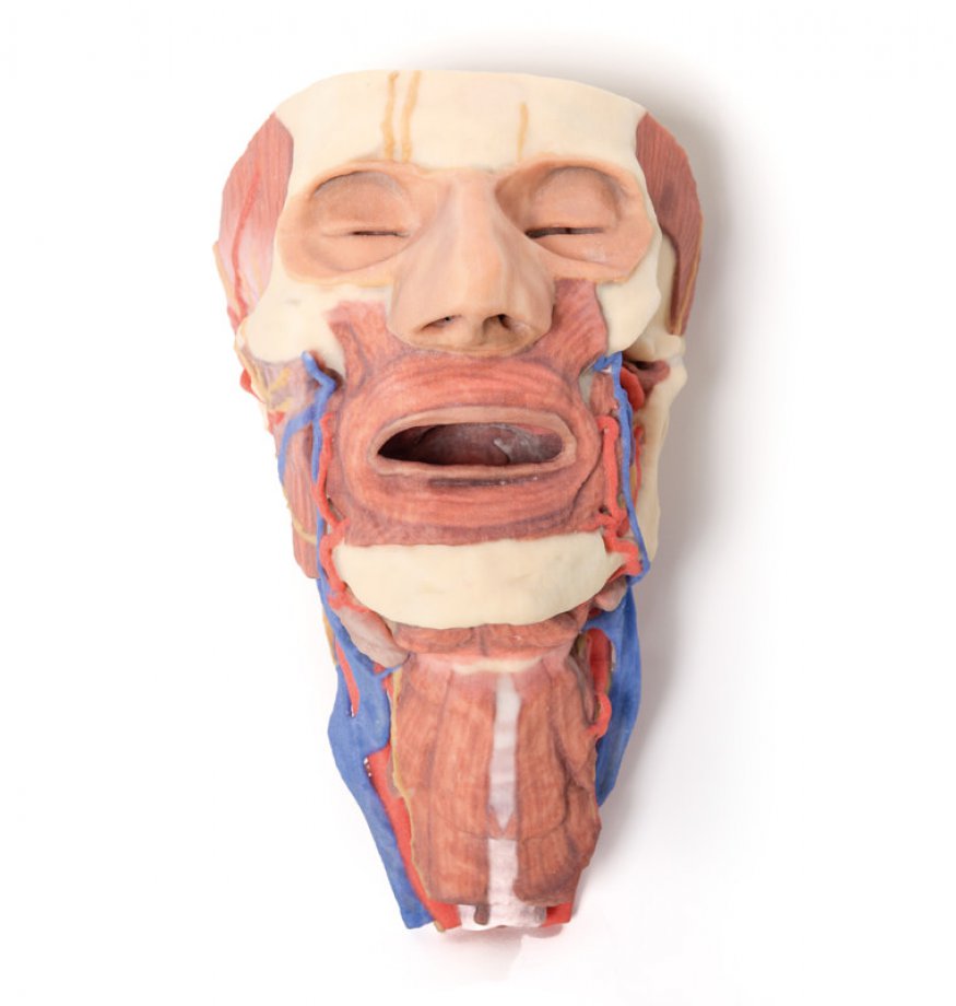
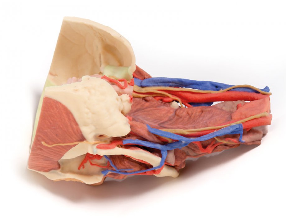
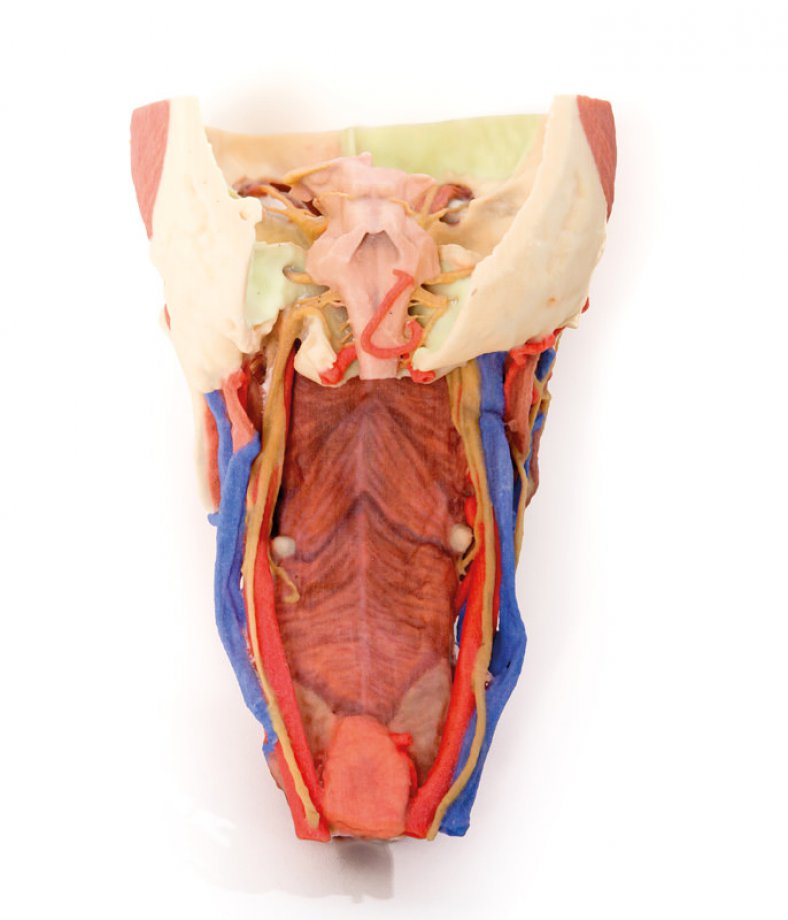
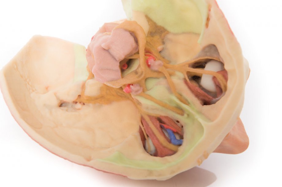
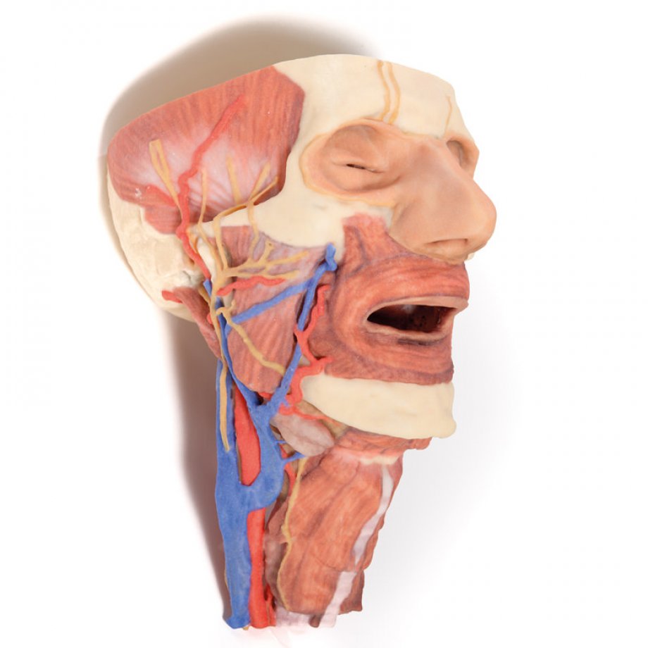
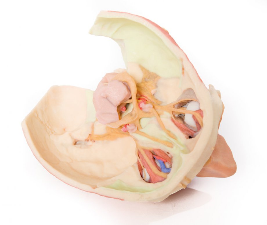
Head and visceral column of the neck
This 3D print specimen preserves a series of features of the head and visceral column of the neck:
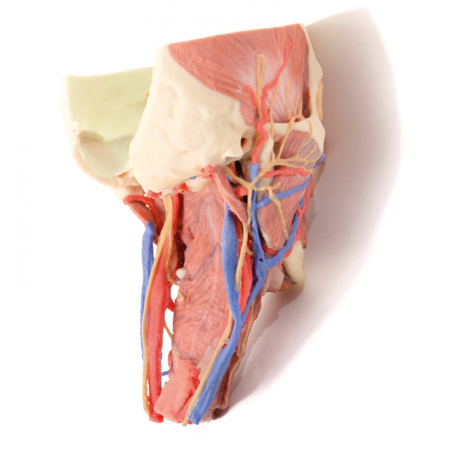
The face: On the right side of the head the parotid gland has been removed to reveal the facial nerve and all its branches (temporal, zygomatic, buccal, marginal mandibular and cervical) and demonstrate the spatial relations of structures embedded in the gland from superficial to deep (facial nerve, retromandibular vein, external carotid artery). In the surrounding region the temporalis, masseter and posterior belly of digastric are exposed, as are and the facial artery, transverse facial artery and superficial temporal artery. The facial vein and transverse facial vein are clearly visible uniting to form the common facial vein which is joined by the retromandibular vein to form the external jugular vein.
Detailed anatomical description on request.