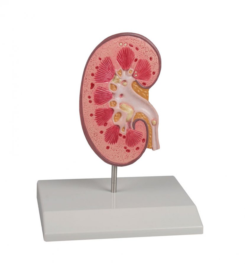
Kidney Stone Model
This model is designed to inform patients about urinary stones (urolithiasis) and kidney stones (nephrolithiasis). A right kidney in natural size is
opened to show the internal structures. The renal pelvis, the renal calices and the ureter are opened to show concretions and stones in the following
locations which are typical:
- origin of the upper calix group
- connecting tubule of the lower calyx group, resulting in congestion of the minor calices
- renal cortex
- ureter
- renal pyramids
Mounted on base. With key card.

